Internal Anatomy & Physiology
Alpheus parvirostris consists of a typical caridean shrimp internal body-plan. The following internal morphology and physiology are generalised from a typical caridean shrimp.
Musculature
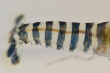
The strongest muscles of Alpheus parvirostris lies not in its enlarged chela but in fact in the pleon (abdomen) (Wicksten 2010). This allows the shrimp to forcefully flip its tail in what is known as a 'tail flip', so that it can make a rapid escape if needs be (Wicksten 2010). Its largest chela consists predominantly of slow contracting muscle in comparison to its smaller chela (pincer) which is made up of intermediate to rapid contracting muscle (Mellon 1999). Physiologically, this makes sense, as the snapping claw is locked into place and therefore needs to stretch significantly for longer periods while the smaller chela would be used for foraging purposing and therefore requires a higher degree of flexibility.
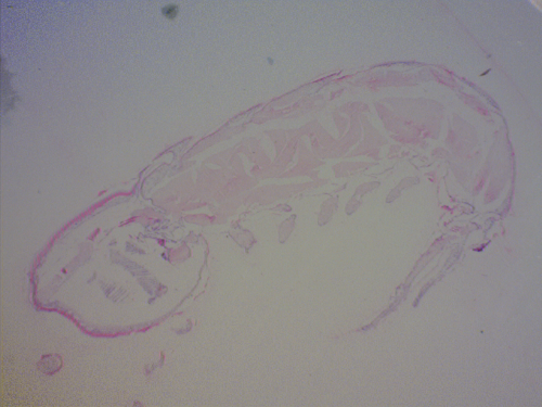
Section slide illustrating musculature and internal anatomy of Alpheus parvirostris. Original Photo Aidan Janetzki 2013.
Circulatory System
The circulatory system of Alpheus parvirostris have what is known as an open circulatory system where organs are bathed directly in oxygen and nutrients rather than via capillaries (Wicksten 2010). Their blood (hemolymph) consists of clotting agents and amebocytes which can aid in immunity and digestion na d a respiratory pigment called hemocyanin (Wicksten 2010). Their heart receives hemolymph via five pairs of ostia and distributes it through numerous anterior and posterior arteries (Wicksten 2010).
Nervous System & Sense Organs
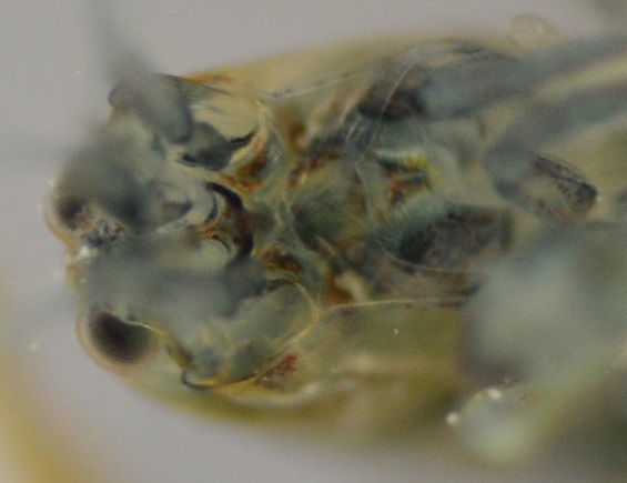
The nervous system of Alpheus parvirostris is based from the cerebral ganglion where optic, antennal and antennular nerves al branch out form (Wicksten 2010). The antennal and antennular nerves run into their respective limbs (see external anatomy). The pleon also possess a major nerve cord, in which five anterior ganglion provide nerves to the musculature and pleopods of the shrimp (Wicksten 2010).
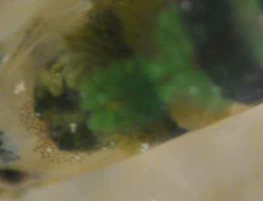
Cerebral ganglion of Alpheus parvirostris (green structure - centre of photo). Original Photo Aidan Janetzki 2013.
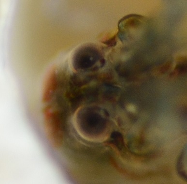
Eyes of Alpheus parvirostris. Original Photo Aidan Janetzki 2013.
Alpheus parvirostris posses two pairs of antennae (second pair named antennule), both of which are composed of tactile receptors (Wicksten 2010). The second pair of antennae however, are also adapted to function in distance chemoreception, in that they are able to sense chemicals within the region (Wicksten 2010). The inner mouth-parts of shrimp also function in chemoreception, but are based off contact only (Wicksten 2010). Snapping shrimp also posses a statocyst, with its primary function involving balance and buoyancy of the shrimp (Wicksten 2010).
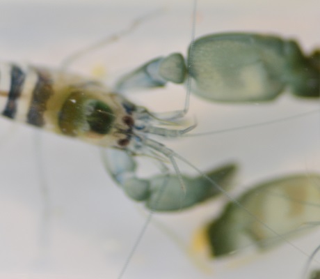
Eyes and antennae of Alpheus parvirostris. Note the two pairs of antennae. Original Photo Aidan Janetzki 2013. Excretory System
The excretory system of Alpheus parvirostris is comprised of antennal glands which empty near the second pair of antennae (Wicksten 2010). Filtration can occur across the epithelium of these structures. Shrimp are predominately osmoconformers, meaning that they will match the internal fluids to the osmolarity of the surrounding seawater. The excretory pore secretes the majority of nitrogenous wastes as ammonia (Wicksten 2010).
Digestive System
The digestive system of Alpheus parvirostris is divided into a two chamber stomach which is connected to the oral opening via a muscualr esophagus (Wicksten 2010). The anterior chamber is known as the cardiac stomach and this is where the crushing and sorting of its diet takes place (Wicksten 2010). The crushing is predominately carried out by the gastric mill, in which food is macerated into a pulp (Wicksten 2010). Once grounded sufficiently, food then moves into the pyloric stomach where food is broken down further by digestive enzymes (Wicksten 2010). Larger food particles are unable to continue because of the presence of numerous setae (Wicksten 2010). Once digested adequately, food can pass through both the midgut and hindgut where it is secreted from the anus (Wicksten 2010).
Feeding appendages of Alpheus parvirostris. Original Photo Aidan Janetzki 2013.
 Alpheus parvirostris midgut which extends almost the
 entire length of the pleon. Original Photo Aidan Janetzki 2013
Respiratory Structures
The prominent respiratory structure of Alpheus parvirostris are gills, more accurately phyllobranchiate (Wicksten 2010). This gill types refers to flat gill lamellae located on both sides of a central axis which is made up of blood vessels carrying hemolymph (Wicksten 2010). Water moves into the gill chamber via the vibrations created from the scaphognathites located on the second maxillae and propels across the gills exchanging exchanging gases with the hemolymph (Wicksten 2010). The water is then swept out via oral cavities (Wicksten 2010).
Genital Structures
Alpheus parvirostris demonstrate gonochorism, in that there are separate male and females (Wicksten 2010). Males posses a pair of testes which produce sperm made up of a de-condensed nucleus and a solitary spike (Wicksten 2010). The spermatophore which is injected into the female during mating is stored in the ampoule (Wicksten 2010). Females have paired tubular ovaries, an oviduct and a female gonopore (Wicksten 2010).
|